Receiving medical imaging results can often be a source of anxiety, especially when terms like “shadows” or “dark spots on X-ray” are mentioned. Many people immediately jump to worst-case scenarios, but the reality is far more nuanced. An X-ray is a powerful diagnostic tool that provides a snapshot of the internal structures of your body, and what appears as a “dark spot” to the untrained eye can mean a multitude of things, ranging from completely normal findings to indicators of underlying conditions.
Understanding the basics of X-ray interpretation can help alleviate some of this apprehension and empower you with knowledge when discussing your results with your healthcare provider. This comprehensive guide will delve into what X-rays show, what constitutes a “dark spot,” common causes in different body parts, and the crucial role of professional interpretation. We’ll explore various scenarios, clarify misconceptions, and outline the typical next steps after such a finding, ensuring you have a clearer picture of what these mysterious dark areas on X-ray images might signify.
Understanding X-Rays and Image Interpretation
Before we dive into dark spots on X-ray images, it’s essential to understand how X-rays work. X-rays are a form of electromagnetic radiation that can pass through the body. Different tissues absorb X-rays to varying degrees:
- Dense tissues like bones absorb more X-rays and appear white on the image.
- Less dense tissues like air (in the lungs) absorb fewer X-rays and appear dark or black.
- Tissues of intermediate density, such as muscle and fat, appear in various shades of gray.
Therefore, a “dark spot” or “lucency” on an X-ray generally indicates an area where X-rays have passed through more easily, suggesting a lower density than the surrounding tissue. This could be due to the presence of air, fluid, fat, or a lack of tissue where it would normally be dense.
The Radiologist’s Role in Interpreting X-Ray Findings
Interpreting X-ray images is a complex skill that requires years of specialized training. Radiologists are medical doctors who specialize in diagnosing and treating injuries and diseases using medical imaging techniques. They meticulously analyze the shades of gray, patterns, and anatomical structures on an X-ray, comparing them to what is considered normal for a patient’s age, sex, and clinical history. What might look like an alarming shadow on X-ray to a layperson could be a normal anatomical variant or a benign finding to an experienced radiologist.
Common Causes of Dark Spots On X Ray Images by Body Area
The significance of a dark spot depends heavily on its location within the body. Let’s explore some common contexts for dark spots on X-ray images.
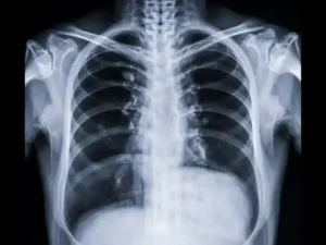
Dark Spots on Chest X-Ray
A chest X-ray is one of the most common imaging tests, often used to evaluate the lungs, heart, and bones of the chest. When a radiologist observes dark spots on X-ray lungs, it typically indicates an area of increased lucency, meaning more air or less dense tissue than expected. Possible causes include:
- Pneumothorax: This is a collapsed lung, where air leaks into the space between the lung and chest wall (pleural space). On an X-ray, this free air appears very dark, pushing the lung away from the chest wall.
- Emphysema or Bullae: These conditions involve damaged, over-inflated air sacs in the lungs, creating abnormally large air spaces that appear darker than healthy lung tissue. Bullae are large air-filled sacs within the lung parenchyma.
- Cysts: Air-filled cysts within the lung can also present as well-defined dark areas.
- Diaphragmatic Hernia: In rare cases, bowel loops (filled with gas) can herniate into the chest cavity, appearing as dark, gas-filled structures.
- Normal Anatomy/Artefacts: Sometimes, overlapping structures or specific patient positioning can create areas that appear darker but are entirely normal.
Dark Spots on Bone X-Ray
When it comes to skeletal imaging, dark spots on X-ray bones typically signify areas where bone tissue has been lost or is less dense than the surrounding healthy bone. These are often referred to as “lytic lesions” or “radiolucent lesions.”
- Osteolytic Lesions: These are areas of bone destruction, where normal bone tissue is replaced by softer tissue, often seen with:
- Bone Tumors: Both benign (e.g., bone cysts, fibrous dysplasia) and malignant (e.g., metastatic cancer, multiple myeloma, primary bone sarcomas) tumors can cause bone destruction.
- Infections (Osteomyelitis): Bacterial or fungal infections can lead to bone inflammation and destruction.
- Bone Cysts: Fluid-filled sacs within the bone.
- Granulomas: Inflammatory lesions.
- Fractures: While a complete break is often a distinct dark line, older or healing fractures might show areas of bone resorption that appear darker.
- Marrow Edema: Although better seen on MRI, severe fluid accumulation in the bone marrow can sometimes present as a subtle lucency on X-ray.
- Normal Variations: Nutrient foramina (small holes where blood vessels enter the bone) or growth plates in children can sometimes be mistaken for lesions.
Dark Spots on Abdominal X-Ray
An abdominal X-ray primarily looks at the bowel gas patterns, kidneys, and surrounding structures. Dark spots on abdominal X-ray usually relate to gas or fluid collections:
- Normal Bowel Gas: The most common “dark spots” are simply gas within the intestines, which is a normal finding. The pattern and distribution of this gas are what radiologists evaluate.
- Abnormal Gas Collections:
- Pneumoperitoneum: Free air in the abdominal cavity, often indicating a perforated organ (e.g., perforated ulcer). This is a serious finding.
- Abscesses: While often appearing as a mixed density, some abscesses can have gas within them, creating dark areas.
- Ileus or Obstruction: Abnormal dilation of bowel loops due to obstruction or paralysis can show exaggerated gas patterns.
- Cysts or Masses: Large cysts or soft tissue masses can sometimes appear darker if they contain fat or are less dense than surrounding structures, but this is less common than in other imaging modalities.
Interpreting What Do Dark Spots On X-Rays Mean: Beyond the Image
It’s vital to remember that an X-ray is just one piece of the diagnostic puzzle. The finding of a dark spot on X-ray never stands alone. A radiologist integrates the imaging findings with several other critical pieces of information:
- Patient’s Medical History: Previous illnesses, surgeries, chronic conditions, and family history can provide crucial context.
- Symptoms: The patient’s current complaints (e.g., pain, fever, shortness of breath) guide the interpretation and narrow down the possibilities.
- Physical Examination: Findings from a doctor’s physical exam are essential for correlation.
- Other Diagnostic Tests: Blood tests, urine tests, or other imaging modalities (like CT scans, MRI, or ultrasound) are often used to further investigate a suspicious finding on an X-ray.
For instance, a small dark spot on a chest X-ray in a young, asymptomatic individual might be dismissed as a normal variant, whereas the same finding in an elderly patient with a history of cancer and new symptoms would warrant immediate and thorough investigation.
When Is a Dark Spot Concerning?
While many X-ray findings of dark spots are benign, certain characteristics can raise concern for radiologists:
- Irregular or ill-defined borders: Suggests aggressive growth.
- Rapid change in size: A spot that grows quickly over time.
- Associated symptoms: Fever, unexplained weight loss, persistent pain, severe shortness of breath.
- Patient risk factors: Smoking history, exposure to certain chemicals, family history of certain diseases.
Next Steps After Identifying Dark Spots on X-Ray
If your X-ray reveals a dark spot that warrants further investigation, your doctor will typically recommend one or more of the following:
- Further Imaging:
- CT Scan (Computed Tomography): Provides much more detailed cross-sectional images, helping to better characterize the size, shape, and density of the dark spot.
- MRI (Magnetic Resonance Imaging): Excellent for soft tissue detail and can differentiate between various tissue types, especially useful for bone marrow or soft tissue lesions.
- Ultrasound: Often used for abdominal or superficial lesions to determine if a dark spot is solid or fluid-filled.
- Blood Tests: To check for markers of inflammation, infection, or specific types of cancer.
- Biopsy: If a solid mass is suspected, a small tissue sample may be taken and examined under a microscope to determine its nature (e.g., benign, malignant, infectious).
- Specialist Consultation: Referral to a pulmonologist for lung issues, an orthopedist for bone issues, or an oncologist if cancer is suspected.
- Observation (Watchful Waiting): For small, indeterminate findings, your doctor might recommend periodic follow-up X-rays to monitor for any changes over time.
FAQ Section: Addressing Your Concerns About Dark Spots On X Ray
Q1: Are all dark spots on X-ray images serious?
A: No, absolutely not. Many dark spots can be normal anatomical variations, benign findings, or even temporary issues like gas in the bowel. The seriousness depends entirely on the location, size, characteristics, and the patient’s overall clinical picture and symptoms. Only a trained medical professional can determine its significance.
Q2: Can a dark area on X-ray disappear on its own?
A: Some can, especially if they are related to transient conditions. For example, a small pneumothorax might resolve spontaneously, or gas patterns in the abdomen can change. However, dark spots caused by structural changes like bone lesions or persistent cysts are unlikely to disappear without intervention.
Q3: What’s the difference between a dark spot and a white spot on an X-ray?
A: On an X-ray, dark areas (radiolucencies) indicate less dense tissue or structures that X-rays pass through easily, like air, fluid, or areas of bone destruction. White areas (radiopacities) indicate denser tissues that absorb more X-rays, such as bones, calcifications, or solid masses.
Q4: If my X-ray shows a dark spot, does it mean I have cancer?
A: Not necessarily. While some cancers can cause dark spots (especially in bone, where they destroy bone tissue), there are numerous other benign causes. It’s crucial not to self-diagnose. Further tests are almost always required to confirm or rule out cancer.
Q5: Should I get a second opinion on my X-ray results?
A: It’s always your right to seek a second opinion, especially if you feel uncertain about your diagnosis or treatment plan, or if the findings are complex. Many institutions encourage second opinions for significant diagnoses to ensure accuracy and peace of mind.
Q6: How quickly should I follow up on a concerning dark spot?
A: The urgency of follow-up depends on what your doctor suspects. If a serious condition is a possibility, they will advise immediate further investigations. For less concerning findings, follow-up might be scheduled for weeks or months later. Always adhere to your doctor’s recommendations for follow-up.
Conclusion
Encountering the term “dark spots on X-ray” can be unsettling, but it’s important to approach such findings with a balanced perspective. These radiological observations are merely pieces of a larger puzzle, indicating areas of lower density within the body. Their meaning can range from completely benign anatomical variations to potential indicators of underlying health conditions that require further investigation.
The key takeaway is the absolute necessity of professional interpretation. Radiologists, with their specialized training, correlate X-ray images with your clinical history and symptoms to provide an accurate assessment. Should a dark spot be identified, remember that it’s the beginning of a diagnostic process, not the end. Your healthcare provider will guide you through the necessary next steps, which may include additional imaging, laboratory tests, or specialist consultations. Stay informed, ask questions, and trust in the expertise of your medical team to unravel the mystery and ensure your best health outcomes.
“`