
If you’ve recently had an eye exam and your doctor mentioned seeing a dark spot on retinal image, it’s natural to feel a bit concerned. Our eyes are incredibly complex organs, and the retina, in particular, plays a crucial role in how we perceive the world. A finding like a dark spot can raise many questions: Is it serious? What caused it? What happens next?
This comprehensive guide aims to shed light on what a dark spot on retinal image means, exploring its various causes, diagnostic procedures, and potential management strategies. From benign pigment changes to more significant medical conditions, we’ll delve into the nuances of this ophthalmic finding. Understanding the possibilities can empower you to have a more informed conversation with your eye care professional and take proactive steps toward maintaining your vision health. Let’s explore the intricate world behind your eyes and decode the message a dark spot on retinal image might be sending.
What is a Dark Spot On Retinal Image? Understanding the Basics
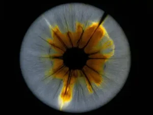
When an ophthalmologist or optometrist examines your eye, they often take images of your retina – the light-sensitive tissue at the back of your eye. These images, known as fundus photographs or OCT scans, provide a detailed view of your retinal health. A dark spot on retinal image refers to an area that appears darker than the surrounding retinal tissue. This can be due to various reasons, ranging from benign anatomical variations to signs of underlying eye conditions.
Anatomy of the Retina and Image Interpretation
The retina is a thin layer of tissue at the back of the eye that contains millions of light-sensitive cells (rods and cones). These cells detect light and send signals to the brain, allowing us to see. Key structures in the retina include the macula (responsible for central vision), the optic disc (where the optic nerve leaves the eye), and blood vessels. When imaging the retina, different tissues and layers absorb or reflect light differently. A dark spot on retinal image can represent:
- An accumulation of pigment.
- An area where light is blocked from reaching the deeper layers.
- A shadow cast by an elevated or abnormal structure.
- An area of tissue atrophy or thinning.
Interpreting these images requires specialized training and equipment, which is why your eye care professional is best equipped to explain any findings.
Common Presentations of a Dark Spot
A dark spot on retinal image isn’t a single entity; it can manifest in several ways:
- Pigmented Lesions: These are often flat, well-defined areas of increased pigmentation, like a freckle in the eye.
- Shadowing: Sometimes, an elevated structure or fluid can cast a shadow, making an area appear darker.
- Hemorrhages: Old or resolving bleeding within the retina can sometimes appear as dark areas.
- Atrophic Areas: Regions where retinal tissue has thinned or degenerated can also present as darker patches due to changes in light reflection.
The size, shape, location, and borders of the dark spot are all crucial details that help your doctor determine its nature.
Why Do Dark Spots Appear on Retinal Images? Potential Causes
The appearance of a dark spot on retinal image can be attributed to a spectrum of causes, from entirely harmless findings to those requiring immediate attention. It’s important to remember that only a qualified eye care professional can accurately diagnose the cause.
Benign Conditions
Many dark spots are benign and pose no threat to your vision. These often include:
- Retinal Pigment Epithelium (RPE) Hyperplasia/Hypertrophy: The RPE is a layer of cells beneath the photoreceptors. Areas where these cells are more numerous or larger can appear as a dark spot on retinal image. These are very common and usually harmless.
- Choroidal Nevus: Similar to a mole on your skin, a choroidal nevus is a benign collection of pigmented cells in the choroid (the vascular layer beneath the retina). Most nevi remain stable and don’t affect vision. However, like skin moles, they need to be monitored for changes.
- Congenital Hypertrophy of the RPE (CHRPE): This is a specific type of benign pigmented lesion, often appearing as a flat, dark, well-demarcated spot, sometimes with a clear halo or small depigmented areas within it. They are typically stable and harmless.
More Serious Conditions
While less common, a dark spot on retinal image can sometimes be a sign of a more serious underlying condition that requires medical intervention. These include:
- Age-related Macular Degeneration (AMD): In some forms of AMD, particularly the “wet” type, blood or fluid leakage can lead to pigment changes or subretinal hemorrhage, which may appear as dark areas.
- Retinal Hemorrhage: Bleeding within the retina or under it can initially appear red, but as it resolves or if it’s deeper, it can present as a darker lesion. Causes include diabetes, high blood pressure, or retinal tears.
- Retinal Detachment: While not a direct cause of a “dark spot” in the typical sense, a retinal detachment can involve areas of hemorrhage or RPE disturbance that might present as darker regions on imaging. Patients often experience acute vision changes, floaters, and flashes.
- Choroidal Melanoma: This is a rare but serious cancerous tumor of the eye. It is the most common primary intraocular malignancy in adults. A choroidal nevus can, in rare instances, transform into a melanoma, making regular monitoring crucial.
- Retinal Scarring: Following an infection, inflammation, or trauma, scar tissue can form on the retina, which may appear darker on imaging.
Artifacts and Imaging Quirks
Sometimes, a “dark spot” might not be a physical finding in the eye at all, but rather an artifact of the imaging process. This could be due to:
- Lens Opacities: Cataracts or other opacities in the eye’s lens can block light, creating shadows on the retinal image.
- Dust or Debris: Particles on the imaging equipment’s lens can also cause dark spots on the image.
- Patient Movement: If the patient moves during imaging, it can lead to distortions that appear as abnormalities.
Your eye care professional will be able to differentiate between true retinal findings and imaging artifacts.
The Diagnostic Process: What to Expect When a Dark Spot is Found
Finding a dark spot on retinal image is the beginning, not the end, of the diagnostic journey. Your ophthalmologist will follow a structured approach to determine its nature and significance.
Initial Examination and Imaging Techniques
Upon detecting a dark spot, your doctor will likely perform a thorough dilated eye examination. This involves using special instruments to look inside your eye. In addition to the initial fundus photography, other advanced imaging techniques might be used:
- Optical Coherence Tomography (OCT): This non-invasive test uses light waves to take cross-sectional images of your retina, allowing the doctor to see the different layers and identify any fluid, swelling, or changes in tissue structure related to the dark spot on retinal image.
- Fundus Autofluorescence (FAF): FAF images highlight lipofuscin, a byproduct of retinal metabolism, which can accumulate in certain retinal diseases or be absent in areas of RPE atrophy. This helps characterize pigmented lesions.
- Fluorescein Angiography (FA): A dye is injected into your bloodstream, which then travels to your eyes. A special camera takes pictures as the dye passes through the retinal blood vessels, revealing any leaks, blockages, or abnormal vessel growth. This is particularly useful for assessing vascular issues or certain tumors.
- Indocyanine Green Angiography (ICG): Similar to FA but uses a different dye that better visualizes the choroidal circulation, which is useful for diagnosing conditions affecting the choroid, such as choroidal nevi or melanomas.
- Ocular Ultrasound: If the dark spot is particularly large or if there’s significant opacity preventing a clear view of the retina, an ultrasound can provide images of the back of the eye and orbital structures.
Differentiating Between Benign and Pathological Findings
The key challenge for your eye doctor is to differentiate between a harmless dark spot on retinal image and one that signals a serious condition. They will consider factors such as:
- Size and Shape: Larger, irregular lesions are often more concerning.
- Color and Pigmentation: Uniformly dark vs. varied pigmentation.
- Presence of Drusen or Fluid: Indicators of macular degeneration or other pathology.
- Associated Symptoms: Changes in vision, floaters, flashes, or pain.
- Growth Over Time: Comparing current images to previous ones is vital. Rapid growth is a red flag.
- Patient’s Medical History: Conditions like diabetes, hypertension, or a family history of eye disease can increase risk.
Based on these findings, your doctor will determine if the dark spot requires observation, further testing, or immediate treatment.
The Importance of Regular Eye Exams
Regular comprehensive eye exams are paramount, especially as you age or if you have risk factors for eye diseases. They allow for early detection of conditions like a dark spot on retinal image and enable timely intervention, which can often prevent significant vision loss. Don’t wait for symptoms to appear; many serious eye conditions progress silently in their early stages.
Treatment and Management of Retinal Dark Spots
The management of a dark spot on retinal image is entirely dependent on its underlying cause. There is no one-size-fits-all treatment, emphasizing the importance of an accurate diagnosis.
When No Treatment is Needed (Observation)
For many benign conditions, such as small choroidal nevi, RPE hyperplasia, or CHRPE, no active treatment is required. Instead, your ophthalmologist will recommend a schedule for regular follow-up examinations, typically every 6 to 12 months. This is crucial for monitoring the dark spot on retinal image for any signs of change or growth, which could indicate a progression to a more serious condition.
- Regular Monitoring: Consistent check-ups with dilated exams and repeat imaging (fundus photography, OCT) are vital.
- Patient Education: Understanding the nature of your specific dark spot and knowing what symptoms to watch for is empowering.
Specific Treatments for Underlying Conditions
If the dark spot on retinal image is a symptom of an underlying disease, treatment will focus on addressing that specific condition:
- Age-related Macular Degeneration (AMD):
- Wet AMD: Injections of anti-VEGF medications (e.g., Avastin, Lucentis, Eylea) into the eye are the primary treatment to stop abnormal blood vessel growth and leakage.
- Dry AMD: While there’s no cure, specific vitamin formulations (AREDS/AREDS2) can slow progression in intermediate stages.
- Retinal Hemorrhage: Treatment depends on the cause. This might involve managing underlying conditions like diabetes or hypertension, laser treatment for leaking blood vessels, or surgery in severe cases.
- Choroidal Melanoma: Treatment options include radiation therapy (brachytherapy), laser therapy (transpupillary thermotherapy), photodynamic therapy, or, in rare cases, surgical removal of the eye (enucleation). Early detection is key for preserving vision and life.
- Retinal Detachment: This is a medical emergency requiring surgical repair, such as vitrectomy, scleral buckle, or pneumatic retinopexy, to reattach the retina.
Lifestyle and Preventative Measures for Retinal Health
While you can’t prevent every cause of a dark spot on retinal image, certain lifestyle choices can significantly contribute to overall retinal health and potentially reduce the risk of some serious conditions:
- Healthy Diet: Consume a diet rich in fruits, vegetables (especially leafy greens), and omega-3 fatty acids (found in fish). These nutrients are vital for retinal function.
- Quit Smoking: Smoking is a major risk factor for several eye diseases, including AMD.
- UV Protection: Wear sunglasses that block 99-100% of UV-A and UV-B radiation when outdoors.
- Manage Systemic Conditions: Keep conditions like diabetes and high blood pressure well-controlled, as they can severely impact retinal health.
- Regular Exercise: Promotes overall cardiovascular health, which benefits eye health.
- Blue Light Protection: While research is ongoing, some eye care professionals recommend blue light filtering lenses for prolonged digital screen use.
Recommended Products for Eye Health Support
While no product can treat a diagnosed retinal condition, certain supplements and protective eyewear can support overall eye health, especially when recommended by your eye care professional. These are general suggestions for maintaining healthy eyes.
AREDS 2 Eye Vitamins
Formulated with specific antioxidants, zinc, lutein, and zeaxanthin, proven to reduce the risk of progression in intermediate Age-related Macular Degeneration (AMD).
Blue Light Blocking Glasses
Help reduce digital eye strain and potential long-term retinal exposure to high-energy visible (HEV) blue light from screens, though direct retinal benefits are still being researched.
Omega-3 Fish Oil Supplements
DHA, a key omega-3 fatty acid, is a major structural component of the retina and is important for maintaining retinal function and overall eye health.
Frequently Asked Questions About Dark Spots on Retinal Images
Q1: Is a dark spot on my retinal image always serious?
A: Not necessarily. While any unusual finding warrants investigation, many dark spots are benign. Conditions like choroidal nevi (eye freckles) or RPE hyperplasia are common and usually harmless. However, it’s crucial to have any dark spot on retinal image evaluated by an ophthalmologist to rule out more serious underlying conditions such as macular degeneration, hemorrhage, or even rare tumors like melanoma.
Q2: What tests will an ophthalmologist perform if they find a dark spot?
A: Beyond a standard dilated eye exam and fundus photography, your ophthalmologist may use advanced imaging. This often includes Optical Coherence Tomography (OCT) to view retinal layers, Fundus Autofluorescence (FAF) to assess RPE health, and sometimes Fluorescein Angiography (FA) or Indocyanine Green (ICG) Angiography to examine blood vessels. Ocular ultrasound might also be used for larger lesions or if views are obscured.
Q3: Can diet affect dark spots on the retina?
A: A healthy diet plays a significant role in overall retinal health, though it may not directly cause or cure all types of “dark spots.” A diet rich in antioxidants, lutein, zeaxanthin (found in leafy greens), and omega-3 fatty acids (from fish) can help protect the retina and reduce the risk of conditions like Age-related Macular Degeneration (AMD), which can sometimes present with pigment changes. Always discuss dietary changes with your doctor.
Q4: How often should I get my eyes checked if I have a dark spot?
A: The frequency of follow-up depends on the nature of the dark spot on retinal image. For benign findings like a choroidal nevus, your doctor might recommend annual or semi-annual check-ups to monitor for any changes. If the spot is suspicious or related to an active condition, more frequent visits, possibly every few weeks or months, may be necessary. Always adhere to your ophthalmologist’s specific recommendations.
Q5: Can dark spots resolve on their own?
A: It depends on the cause. Benign pigmented lesions like nevi typically remain stable and do not resolve. If a dark spot is due to a resolving hemorrhage, it may fade over time as the blood is reabsorbed. However, dark spots caused by permanent tissue damage, scarring, or certain degenerative diseases will not typically resolve on their own. It’s essential for your eye doctor to monitor and manage the underlying condition.
Conclusion
Discovering a dark spot on retinal image can be a source of anxiety, but it’s crucial to approach this finding with informed understanding rather than immediate panic. As we’ve explored, these spots represent a diverse range of conditions, from entirely benign and harmless variations in retinal pigmentation to more serious indicators of underlying eye disease. The key takeaway is that such a finding always warrants a comprehensive evaluation by a qualified ophthalmologist.
Your eye doctor will utilize advanced imaging and their expertise to accurately diagnose the nature of the dark spot, differentiate between benign and pathological causes, and recommend an appropriate course of action, be it careful observation or specific treatment. Remember, early detection and regular eye examinations are your best defense against potential vision loss. By staying proactive about your eye health and maintaining open communication with your eye care professional, you can ensure the best possible outcomes for your precious vision.